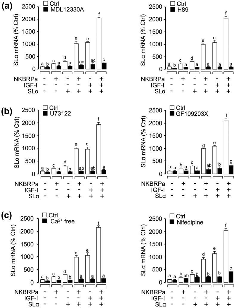
Figure 5. Functional role of AC/PKA, PLC/PKC and Ca2+ -dependent signaling pathways in the regulation of SLα mRNA expression.
In this experiment, carp pituitary cells were incubated for 24 hours in the absence (control) or presence of (a) AC inhibitor MDL12330A (10 μM) and PKA inhibitor H89 (10 μM), (b) PLC inhibitor U73122 (10 μM) and PKC inhibitor (10 μM), or (c) Ca2+ free medium and VSCC blocker nifedipine (10 μM) in combination of NKBRPa (1 μM), SLα (30 nM), SLα (30 nM)+ NKBRPa (1 μM), SLα (30 nM)+ IGF-I (50 nM), or SLα (30 nM)+ NKBRPa (1 μM)+ IGF-I (50 nM). After drug treatment, total RNA was isolated for real-time PCR of SLα mRNA. In the data present (mean ± SEM), the groups denoted by different letters represent a significant difference at p < 0.05 (ANOVA followed by Dunnett’s test).

