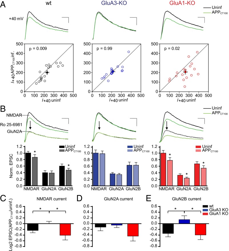
Fig. 2.
GluA3-deficient neurons are resistant against Aβ-mediated synaptic NMDAR depression as shown by dual whole-cell recordings of APPCT100-infected and neighboring uninfected CA1 neurons in organotypic slices from WT mice (black), GluA3-KO littermate mice (blue), or GluA1-KO littermate mice (red). (A) Sample traces (Upper) and dot plots (Lower) of paired NMDAR EPSC recordings (open dots) with the averages shown as filled dots (WT, n = 17; GluA3-KO, n = 16; GluA1-KO, n = 17). Genotype × APPCT100: P = 0.05 (two-way ANOVA). (Scale bars: 20 ms and 50 pA.) (B) Sample traces (Upper) and average EPSC currents normalized to the average of the uninfected neurons (Lower) before and after Ro 25-6981 wash-in to reveal GluN2A- and GluN2B-contributing NMDAR currents. (Scale bars: 20 ms and 50 pA.) (C–E) Fold change in total NMDAR (C), GluN2A (D), and GluN2B (E) currents upon APPCT100 expression, calculated as the average log2-transformed ratio of EPSCs recorded from APPCT100-infected neurons over EPSCs from neighboring uninfected neurons. Data are mean ± SEM. Statistics: two-tailed paired (A and B) or unpaired (C–E) t test. *P < 0.05.

