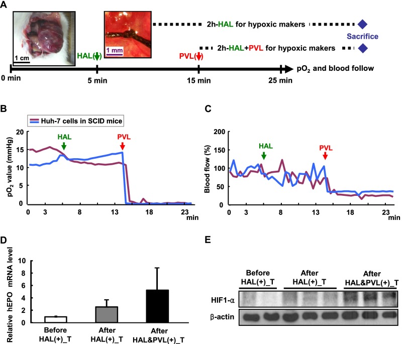
Fig. S1.
Orthotopic xenograft mouse HCC model is supplied by both the HA and PV. (A) Schematic illustration of the pO2 and blood flow measurement process by HAL and PVL in the left liver lobe. The black arrow indicates the combined OxyLite/OxyFlo probes inserted into the hepatic tumor to monitor the pO2 and blood flow continuously. The green arrow indicates the time of HAL, and red arrow indicates the time of PVL. The blue diamond indicates the sacrifice point. (B and C) Hypoxia cannot be established in the hepatic tumor of the orthotopic xenograft mouse model when only HAL was used. The probes were inserted into the hepatic tumor to monitor the pO2 (B) and blood flow (C) continuously. (D and E) Hypoxic markers of EPO and HIF1-α cannot be induced in the hepatic tumor of the orthotopic xenograft mouse model when only HAL was used for 2 h. Tumor tissues were harvested before, after the 2-h HAL, and after the 2-h combined HAL and PVL treatments from the left liver lobe. All samples were evaluated for the EPO mRNA by quantitative RT-PCR (D) and HIF-1α protein levels (E) by Western blotting, with the tumor of left liver lobe before L-HAL as control.

