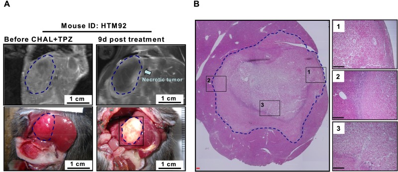
Fig. S9.
Contrast-enhanced MRI can detect the necrosis in HCC caused by TPZ combined with transient CHAL. (A) The MRI images and the gross appearance of an HCC in liver (marked in blue dashed circles), at time points before treatment (Left) and at 9 d after treatment (Right). (B) The liver tissues were collected for H&E staining, with the results shown in a manner covering the whole cross-section of the tumor region to support the near-complete necrosis after treatment. Several areas at the boundary or central parts of the tumor marked by black dashed squares were demonstrated in a higher magnification manner in Insets. (Scale bars: 0.2 mm.)

