Fig. 2.
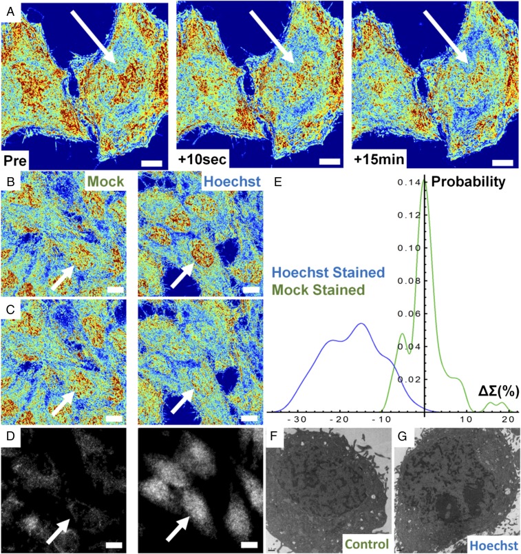
Hoechst excitation induces rapid transformation of chromatin nanoarchitecture. (A) Pseudocolored live-cell PWS image of Hoechst 33342-stained HeLa cells before and after excitation of the dye with UV light. Transformation of chromatin occurs across the whole nucleus within seconds and no repair is observed even after 15 min. (B) Hoechst-stained and M-S cells before excitation and (C) the same M-S and Hoechst-stained cells after UV irradiation. (D) Minimal (mock) and significant (Hoechst) γH2A.X antibody accumulation. (E) Distribution of chromatin transformation after UV excitation for Hoechst-stained and M-S cells. (F and G) Transmission electron-microscopic images of control and Hoechst-stained cells confirming nanoscale fragmentation of the chromatin nanoarchitecture in fixed cells. All pseudocolored images scaled between Σ = 0.01 and 0.065. (All scale bars: 15 µm.) Arrows indicate representative nuclei.

