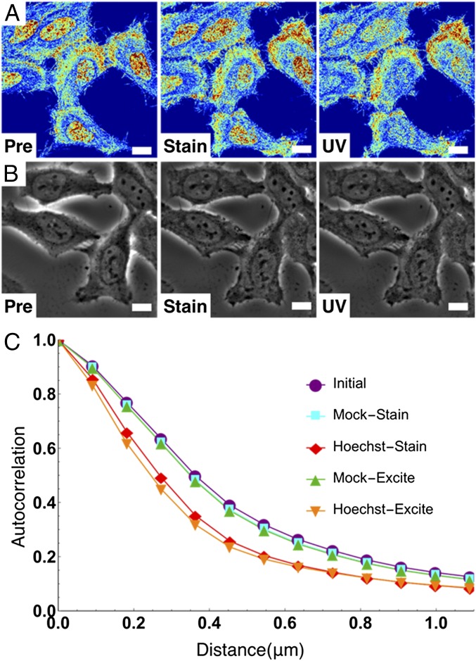
Fig. 3.
Live-cell PWS uniquely detects nanoarchitectural transformation resulting from Hoechst incubation and excitation. (A and B) Live-cell PWS (A) and phase contrast (B) cells preincubation, 15-min postincubation, Hoechst fluorescent image, and after excitation. (C) Change in the autocorrelation function of live-cell PWS intensity. Hoechst transforms chromatin into a more globally heterogeneous structure. Live-cell PWS images are scaled between Σ = 0.01 and 0.065. (All scale bars: 15 µm.)

