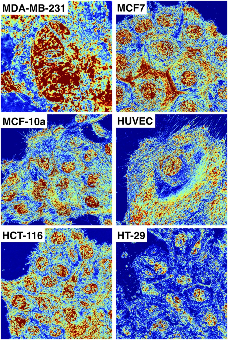
Fig. S5.
Full spectral acquisition of live-cell PWS for six cell lines. Live-cell PWS microscopy allows direct analysis of the nanoscopic topology of a wide range of eukaryotic cell line models. In addition to the ubiquitously used cell lines used in the main manuscript (HeLa and CHO cells), additional classical models of breast cancer (MDA-MB-231, MCF-7, MCF-10a), colon (HT-29, HCT116), and even primary human cell lines (HUVECs) can all be imaged without the need for fluorescent transfection or small-molecule exogenous dyes. ∑ scaled between 0.01 and 0.065 in all cells except MDA-MB-231, which is called from 0.01 to 0.1.

