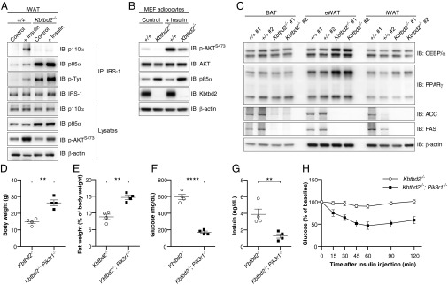
Fig. 6.
p85α accumulation impairs PI3K signaling and causes the tny phenotype. (A) Immunoblot analysis of immunoprecipitates and lysates of iWAT before (Control, anesthesia alone) or 5–6 min after i.v. injection of insulin (+Insulin). (B) Immunoblots of lysates of 10-d-differentiated MEF-derived adipocytes. (C) Immunoblots of lysates of different adipose tissues from two 8-wk-old male Kbtbd2−/− mice or WT littermates. (D–G) Body weight (D), normalized fat weight (E), blood glucose levels (F), and serum insulin levels (G) of 11-wk-old male Kbtbd2−/− and Kbtbd2−/−; Pik3r1−/− mice. Glucose and insulin were measured after a 6-h fast. (H) Insulin tolerance test. Blood glucose was measured at indicated times after i.p. insulin injection in male Kbtbd2−/− mice (n = 4) and Kbtbd2−/−; Pik3r1−/− mice (n = 4) at 11 wk of age. The baseline blood glucose levels (0 min) of Kbtbd2−/− mice and Kbtbd2−/−; Pik3r1−/− mice were 594 ± 32 mg/dL and 172 ± 14 mg/dL, respectively. In D–G, data points represent individual mice. P values were determined by Student’s t test.

