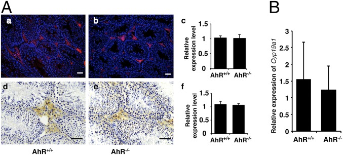
Fig. 1.
AhR depletion did not affect the expression of aromatase. (A) Aromatase protein level remained unchanged in AhR null mouse testis. (A, a and b) IF staining of aromatase protein (red) in AhR+/+ and AhR−/− testis. Slides have been counterstained with DAPI (blue) to mark nuclei. (A, c) Quantification of IF staining using ImageJ software showed that there is no significant difference of aromatase expression between AhR+/+ and AhR−/− mice. (A, d and e) IHC staining of aromatase protein (brown stain) in AhR+/+ and AhR−/− testis. (A, f) Quantification of IHC staining using ImageJ software showed that there is no significant difference of aromatase expression between AhR+/+ and AhR−/− mice. (Scale bars: 25 μm.) (B) AhR depletion does not affect the expression of aromatase gene in male mouse mammary gland. Real-time PCR showed the relative expression of Cyp19a1 in AhR+/+ and AhR−/− mammary glands. The difference is not significant.

