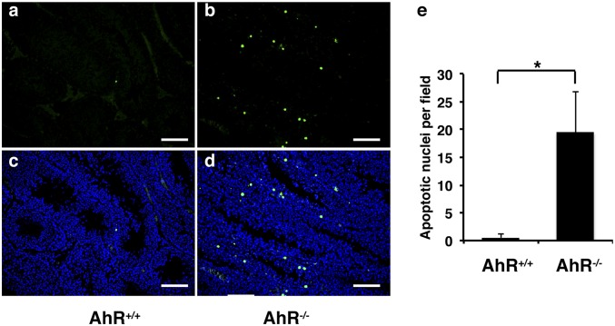
Fig. 4.
TUNEL assay (green fluorescence) to show that there is increased apoptosis in AhR−/− mouse testis. (A and C) AhR+/+ testis. (B and D) AhR−/− testis. (E) Graphic diagram to show the numbers of apoptotic nuclei per field by using 10× lens (*P < 0.01). A and B, TUNEL staining; C and D, DAPI plus TUNEL staining. For counting these apoptotic nuclei, randomly selected stained slides from three mice in each group and more than six representative fields of each slide were counted under light microscope using 10× lens.

