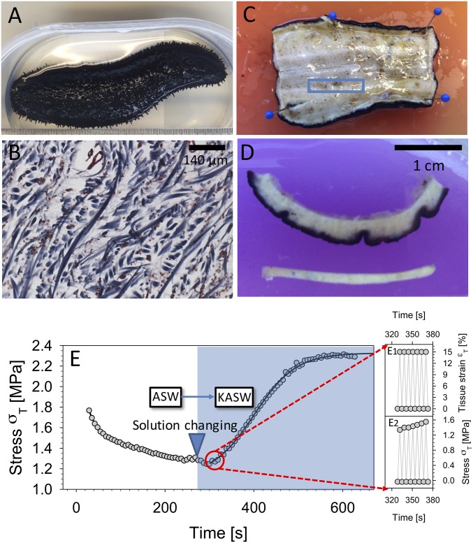
Fig. 1.
Sea cucumber body wall MCT. (A) Sea cucumber H. leucospilota. (B) Transverse section of sea cucumber body wall stained using Masson’s trichrome method. Collagen fibrils appear blue. (C) The sheet of body wall after the animal was cut in half along the longitudinal plane. The blue rectangle indicates the dimensions and location of the sectioned specimen, with the long dimension along the longitudinal axis. (D, Upper) View of sectioned sea cucumber dermis including dark outer dermis and inner layer. (D, Lower) The tensile test specimen. Before testing, the dark-pigmented outer dermis as well as the inner layer was removed, leaving only the center part of the specimen. (E) Time-dependent change in sea cucumber MCT mechanics induced via ionic treatment. The peak stress (per cycle) is plotted during strain-controlled cyclic loading of sea cucumber dermis at 0.3 Hz (to 15% tissue strain), with tissue immersed in ASW until ∼290 s, followed by a change of the immersing solution to KASW (stiffening agent). A clear rise of peak stress (per cycle) is observed, fitted with a sigmoidal curve as a guide to the eye. Insets (E, Right) show a magnified time range over a few (seven) cycles, with both maximum and minimum stress and strain indicated.

