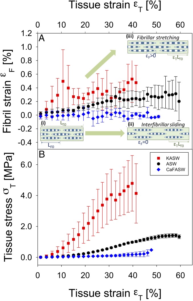
Fig. 3.
Altered fibrillar stress and strain take-up in ionically treated MCT. (A) Fibril strain vs. applied tissue strain from ionically treated sections of MCT dermis, measured from the peak shifts of the fifth-order collagen reflections in the SAXD pattern. The rate of increase of fibril strain with tissue strain (εF/εT) is proportional to the amount of stress taken up by the collagen fibrils. Data are from control (ASW) (black circles, n = 4), stiffened (KASW) (red squares, n = 4), and softened (CaF-ASW) (blue diamonds, n = 3) MCT. All samples in each group are binned according to tissue strain with bin widths of 2.0%; error bars are SDs. Stiffened MCT exhibits a higher rate of increase of fibril strain compared with control, whereas softened MCT shows essentially no increase in fibril strain. Inset schematics: (A, i) Fibrils (striated ellipsoids; length LF0) separated by interfibrillar matrix, in unloaded MCT of length LT0; (A, ii) in softened MCT (CaF-ASW treated), while the tissue elongates (εT > 0), the fibrils do not stretch, but slide in the interfibrillar matrix (εF = 0); (A, iii) in stiffened MCT (KASW treated), there is increased stress transfer to the fibrils, leading to fibrillar stretching (εF > 0). (B) Corresponding macroscopic tensile stress/strain curves for the control, stiffened, and softened groups, binned according to tissue strain (error bars: SDs), showing clear differences in tangent modulus and maximum stress achieved.

