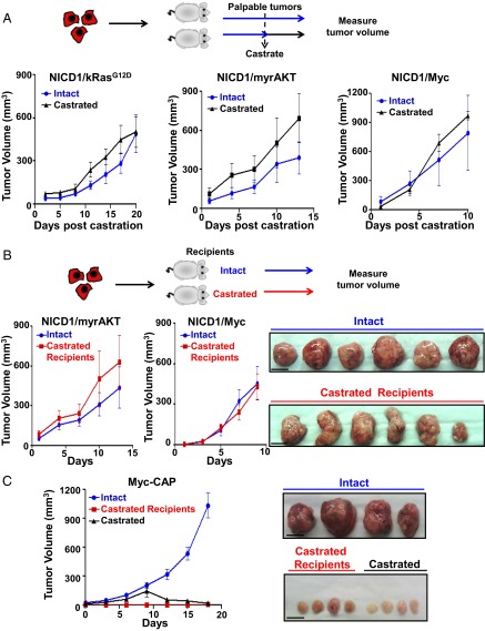
Fig. 6.
NICD1 in combination with the Ras/Raf/MAPK, myrAKT, or Myc pathways drives CRPC. (A, Upper) Schematic representation of the experimental design. Primary tumors driven by NICD1/kRasG12D, NICD1/myrAKT, or NICD1/Myc were dissociated into single cells. NICD1/kRasG12D, NICD1/myrAKT, or NICD1/Myc tumor cells (6 × 10⁵) were mixed with Matrigel and transplanted s.c. into six to eight mice per condition. Upon detection of palpable tumors (50 mm3), six to eight mice carrying a total of six to eight tumors were castrated (marked as day 1). Tumor length, width, and height were measured every 3 d for a total of 10–20 d. (Lower) Tumor volumes were calculated by multiplying length × width × height/2 and were plotted. One of the two independent experiments for each tumor type (NICD1/kRasG12D, NICD1/myrAKT, or NICD1/Myc) is shown. (B, Upper) The experimental design. NICD1/myrAKT or NICD1/Myc tumor cells (6 × 10⁵) were mixed with Matrigel and transplanted s.c. into six castrated or six intact recipient mice. (Lower Left) Upon detection of palpable tumors (50 mm3), marked as day 1, tumor volumes were measured every 3 d. (Lower Right) Recovered NICD1/Myc secondary tumors from intact or castrated recipients are shown. (Scale bars: 1 cm.) One of the two independent experiments for each tumor type (NICD1/myrAKT or NICD1/Myc) is presented. (C, Left) Myc-CAP cells (6 × 10⁵) were mixed with Matrigel and implanted s.c. into eight intact or four castrated immunodeficient recipient mice. Upon detection of palpable tumors, one group of intact mice (n = 4) was subjected to surgical castration. (Right) Images of the recovered tumors. (Scale bars: 1 cm.)

