
Fig. 3.
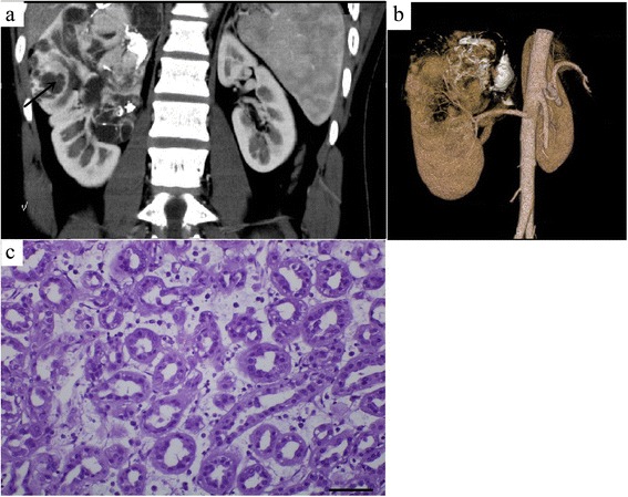
A 17-year-old male with metanephric adenoma in the mid and upper poles of the right kidney (case 2). Dynamic contrast-enhanced CT revealed an irregular lesion with a heterogeneous enhancement, multiple patchy calcifications, and cystic changes/necrosis (a). CT angiography revealed the nutrient artery in the lesion (b). Pathology was assessed with hematein-eosin staining and showed that the morphology of tumor cells was uniform with tubular and acinar architecture (magnification, 40 × 10, c). The arrow marker was used to indicate the cystic changes or necrosis