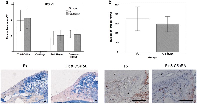
Fig. 3.

a Histological evaluation of the fracture callus for both experimental groups after 21 days with representative histological images of the callus (Fx = fracture; Fx & C5aRA = fracture with administration of C5aRA). b Results of histological evaluation of PMN after 3 days with representative histological images of the callus region in detail. Results are displayed as mean ± 2 × SEM; size of the scale bar = 100 µm; * = cortex, # = periosteal callus
