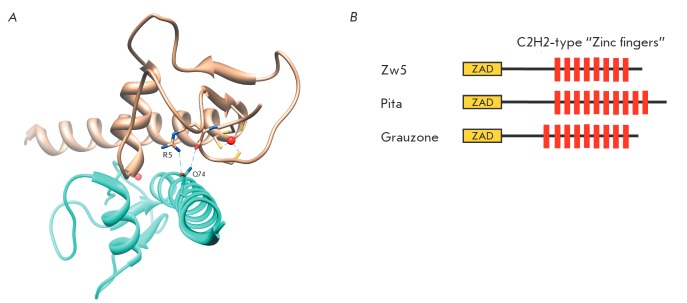
Fig. 2.
A – Crystal structure of ZAD-domain dimer from Grau protein (1PZW in PDB). Hydrogen bonds between R5 (residue homological to R14 of Zeste-white 5 protein) and Q74 are shown; also shown are four cysteines coordinating the zinc-ion. B – Domain structure of the Zeste-white 5, Pita, and Grauzone proteins.

