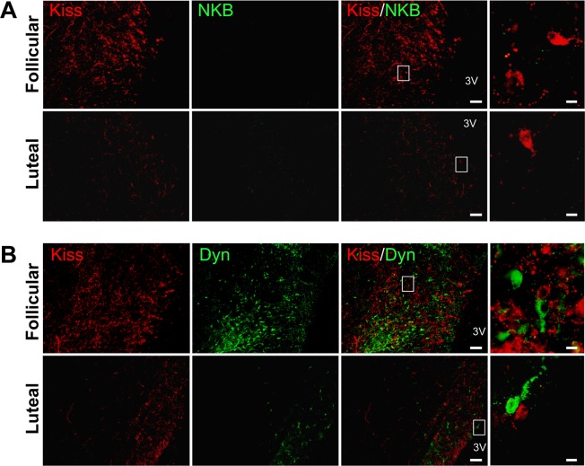
Fig. 3.
Expression of kisspeptin, neurokinin B (NKB), and dynorphin A (Dyn) in the preoptic area (POA) in cows. (A) Representative photomicrographs showing dual immunohistochemistry for kisspeptin (red) and NKB (green) during the follicular and luteal phases. The merged images show the absence of co-localization of kisspeptin and NKB in the POA. (B) Representative photomicrographs showing dual immunohistochemistry for kisspeptin (red) and Dyn (green) during the follicular and luteal phases. The merged images show the absence of co-localization of kisspeptin and Dyn in the POA. The white boxes in the merged images show the areas of the magnified images. Scale bars are 100 μm and 10 μm in the merged and magnified images, respectively. 3V, third ventricle.

