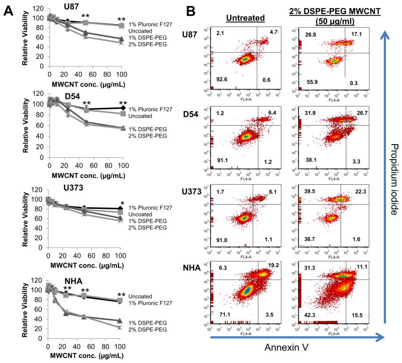
Figure 2. Evaluation of the cytotoxicity induced by MWCNTs modified by acid oxidation and coating with various surfactants.
A) U87, U373, and D54 GBM cells or NHA cells were treated for 24 h with increasing doses of acid oxidized MWCNTs dispersed in the indicated surfactants. After 24 h, cells were lysed, pelleted and the supernatant was analyzed for ATP content as a measure of cell viability using the CellTiter-Glo assay. Samples were prepared and measured in sextuplicate and are displayed as the mean ± standard deviation of each measurement. Significant differences in viability between cells treated with 1% or 2% DSPE-PEG coated MWCNTs and uncoated or Pluronic F-127 coated MWCNTs (determined by ANOVA followed by Student’s T-Test when appropriate) are indicated by (*; p<0.05) or (**; p<0.01). B) U87, U373, and D54 GBM cells or NHA cells were treated for 24 h with 2% DSPE-PEG MWCNTs (50 μg/ml). Cells were co-stained with propidium iodide and Annexin V and then evaluated by flow cytometry. the percentages of cells characterized as viable (lower-left quadrant), early apoptotic (lower-right quadrant), late-apoptotic (upper-right quadrant), and necrotic (upper-left quadrant) are shown within each quadrant. At least 10,000 cells were counted for each measurement and the experiments were repeated 2–3 times for each cell line with similar results.

