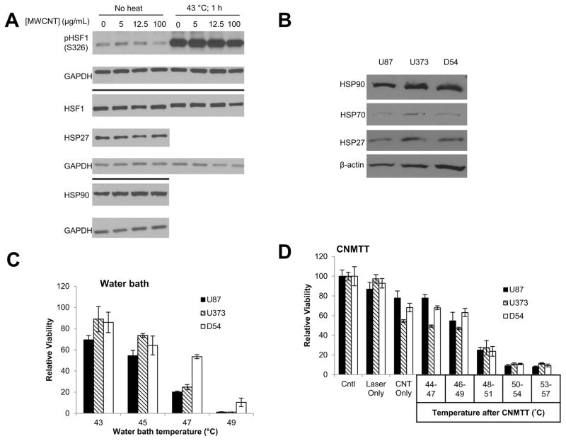
Figure 3. Quantification of heat shock protein expression and response of GBM cells to thermal therapies.
A) Adherent U87 cells were treated with increasing concentrations of 2% DSPE-PEG MWCNTs overnight. To determine if the nanotubes influenced activation of the heat shock response, half of the cells were subsequently exposed to 43 ºC for 1 h. Whole cell lysates were collected and probed for proteins involved in the heat shock response via western blot analysis. GAPDH was used as a loading control. B) Basal expression of heat shock proteins in U87, U373, and D54 GBM cells was assessed by western blot analysis and normalized to β-actin. C) GBM cells were heated to increasing temperatures (43–49 ºC) in a circulating water bath to mimic conventional heating methods. Cells were allowed to recover for two days and cell viability was determined by MTT assay. D) GBM cells were heated to increasing temperatures (44–57 ºC) using CNMTT. Cells were allowed to recover for two days and cell viability was determined by MTT assay. For (C) and (D), data are expressed as the mean of triplicate samples and are normalized to the unheated control for each cell line. Significant decreases in viability with each increase in temperature (p<0.05) were detected for all cell lines. The results shown are representative of at least 3 independent experiments.

