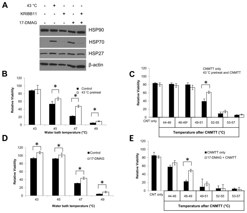
Figure 4. Determination of the effect of sublethal heat or chemically induced heat shock protein expression on thermotolerance of GBM cells.
To induce the HSR, U87 cells were placed in a 43 ºC incubator for 1 hour and recovered at 37 ºC for 4 hours, or treated with a combination of 1 μM 17-DMAG and 10 μM KRIBB11 for 5 hours. KRIBB11 treatment occurred 30 minutes prior to 17-DMAG treatment to allow sufficient time for the inhibitor to take effect. A) Whole cell lysates were collected from all treatment groups and probed for HSP90, HSP70, or HSP27 via western blot analysis. β-actin was used as a loading control. The HSR was induced in U87 cells by placement in a 43 ºC incubator for 1 h and recovery at 37 ºC for 4 h as above. Cells were subsequently heated to increasing temperatures in B) a circulating water bath to mimic conventional heating methods or C) by CNMTT. The HSR was induced in U87 cells by treatment with 1 μM of 17-DMAG for 5 h as above. Cells were subsequently heated to increasing temperatures in D) a circulating water bath E) or by CNMTT. Cells were allowed to recover for two days after each treatment and cell viability was determined by MTT assay. Data are expressed as the mean of triplicate samples normalized to untreated (no heat and no 17-DMAG) controls. Significant differences (p<0.05) between treatment groups determined by Student’s T-Test are indicated by (*). The results shown are representative of 2–3 independent experiments per condition tested.

