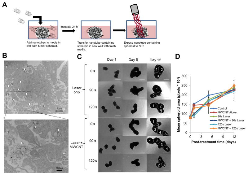
Figure 5. Imaging the diffusion of 2% DSPE-PEG MWCNTs into three dimensional GBM tumor spheroids and treatment of nanotube containing spheroids by CNMTT.
Briefly, U87 cells were grown as three dimensional tumor spheroids and incubated overnight with 2% DSPE-PEG MWCNTs. The spheroids were washed, and then transferred to new wells for use in electron microscopy or photothermal treatment studies. A) Schematic illustrating the experimental design. U87 spheroids incubated overnight with 2% DSPE-PEG MWCNTs were washed, fixed, embedded, sectioned, overlaid onto copper-coated formvar grids and imaged by TEM. In the electron micrographs, B) MWCNTs (indicated by the white arrows) can be seen penetrating throughout the spheroids (upper panel). A higher powered image (lower panel) of the highlighted area indicates that the MWCNTs are both in intracellular compartments and in the extracellular space of spheroids. Groups of 4–6 spheroids alone or spheroids incubated overnight with 2% DSPE-PEG MWCNTs then washed to remove excess nanoparticles were exposed to laser emitted NIR energy (3 W/cm2) for 0–120 s. Spheroid growth over time was monitored and C) representative photomicrographs are shown. Individual spheroids fused into a single cluster are identified in the day 15 images. D) Mean surface area per spheroid was quantified in pixels using Image J software. For area measurements, images of spheroids in addition to those shown in B) were used. No significant differences in spheroid grow were detected between treatment groups.

