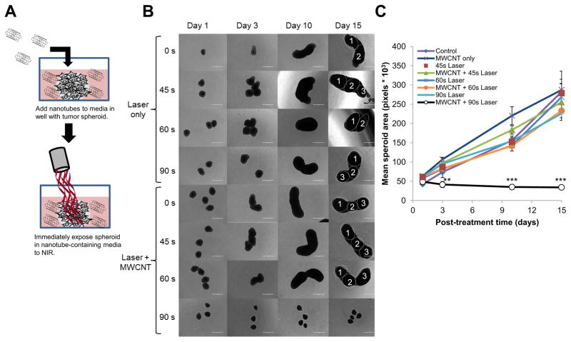
Figure 6. Treatment of GBM spheroids by CNMTT in the presence of extracellular 2% DSPE-PEG MWCNTs.
Briefly, U87 cells were grown as three dimensional tumor spheroids. Groups of 4–6 spheroids were transferred to new wells containing 2% DSPE-PEG MWCNTs (20 μg/ml) in the media, and were exposed to laser emitted NIR energy (3 W/cm2) for 0–90 s. After treatment, the spheroids were washed, and then transferred to new wells with growth media only. A) Schematic illustrating the experimental design. Spheroid growth over time was monitored and B) representative photomicrographs are shown. Individual spheroids fused into a single cluster are identified in the day 15 images. C) Mean surface area per spheroid was quantified in pixels using Image J software. For area measurements, images of spheroids in addition to those shown in B) were used. Significant differences in spheroid grow were detected between spheroids treated with MWCNTs and laser for 90 s as compared to all other treatment groups (determined by ANOVA followed by Student’s T-Test when appropriate) are indicated by (**; p<0.01) or (***; p<0.001).

