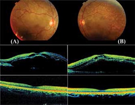
Figure 2. Case 2, postpartum day 5 colored fundus photograph from severe preeclamptic patient showing serous retinal detachment accompanied by many yellow, opaque lesions in the retinal pigment epithelium. Elevation of the inner and outer retinal layers associated with severe neurosensorial retinal detachment and intraretinal cysts were noted in optical coherence tomography images taken at the same time. Complete resolution of the macular edema and normal foveal contours were observed on follow-up optical coherence tomography at 1 year.

