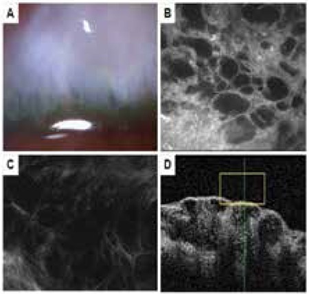
Figure 3. Microcystic bleb. A) Biomicroscopic view of a bleb with microcysts and pronounced elevation above the scleral bed. B) Many fluid-filled, hyporeflective microcysts in the conjunctival epithelium. C) In vivo confocal microscopy image showing loose (grade 0) subepithelial connective tissue. D) Anterior segment optic coherence tomography of the sagital cross-section of the bleb showing many fluid-filled subepithelial and subconjunctival spaces.