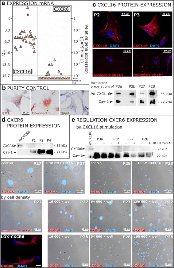
Fig. 1.

Expression of CXCL16 and its receptor CXCR6 in cells cultivated from human meningiomas. a Transcription of CXCL16 and its receptor CXCR6 in human meningioma primary cultures was determined by qRT-PCR (each triangle indicates an individual patient’s sample). Meningioma primary cultures show high mRNA levels of CXCL16, whereas the expression of CXCR6 is hardly detectable or completely lost. b The identity and purity of meningioma cultures was assured for every culture by immunocytochemistry on EMA, fibronectin and GFAP, only cultures with positive reactivity for EMA and fibronectin and negative staining for GFAP were used for further experiments (exemplary data). c Expression of surface tm-CXCL16 on protein level was confirmed by immunocytochemistry using a CXCL16 specific antibody and a red labeled secondary antibody, nuclei were counterstained with DAPI. Furthermore, membrane expression of CXCL16 was proven by western blot using membrane preparations isolated from meningioma cells. Caveolin-1 (Cav-1) served as membrane-specific loading control. d Lack of CXCR6 expression was confirmed on protein level by western blot. In parallel to subsequent experiments, cell lysates were analyzed using a CXCR6-specific antibody. Whereas the positive control, human recombinant CXCR6, was sensitively detected (50 ng/lane), cultured meningioma cells did not show any CXCR6 expression. e CXCR6 expression was neither induced by CXCL16 stimulation nor by different cell densities. Stimulation with 10 nM recombinant human CXCL16 for 24 h, the maximum stimulation time for subsequent experiments, did not yield an induction of CXCR6 expression as shown by ICC and western blot. Additionally, different cell densities in a range from 16,000 to 160,000 cell/well (representing the cell densities seeded for subsequent experiments) did not influence on the expression of CXCR6 as shown by ICC. Exemplary data of technical (P3 a and b different cultivation passages) and biological replicates are shown. LOX melanoma cells which were transfected with an expression vector for human CXCR6 served as a positive control to confirm the specificity of the CXCR6 antibody in ICC, recombinant human CXCR6 served as positive control for western blot experiments
