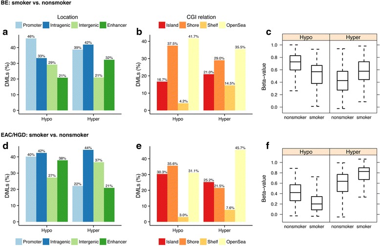
Fig. 5.

Genomic location, relationship to CpG islands, and methylation status of DML when comparing smokers and nonsmokers in esophageal samples. “Hypo” refers to percentage of DML that are hypomethylated in smokers vs. nonsmokers; “Hyper” refers to percentage of DML that are hypermethylated in smokers vs. nonsmokers. On the Y axis, DMLs (%) refers to the percentage of the total DML that are associated with a particular genomic location (a, d) or CGI relationship (b, e). Percentages may up to more than 100 % because some probes were classified with more than one designation. Beta values are equivalent to percent methylation. Note: for all regions, the distribution of hypo/hypermethylated DML compared to the expected distribution (based on all array probes) was not statistically significant. a DML when comparing smoker to nonsmoker BE cases by genomic region. b Location of DML when comparing smoker to nonsmoker BE cases with respect to CpG island location. c Box and whisker plots showing distribution of DML that are hypomethylated in the smoker vs. nonsmoker BE cases (left) and hypermethylated in the smoker vs. nonsmoker BE cases (right). d DML when comparing smoker vs. nonsmoker HGD/EAC cases by genomic region. e Location of DML when comparing smokers vs. nonsmoker HGD/EAC cases with respect to CpG island location. f Box and whisker plots showing distribution of DML that are hypomethylated in the smoker vs. nonsmoker BMI HGD/EAC cases (left) and hypermethylated in the smoker vs. nonsmoker HGD/EAC cases (right)
