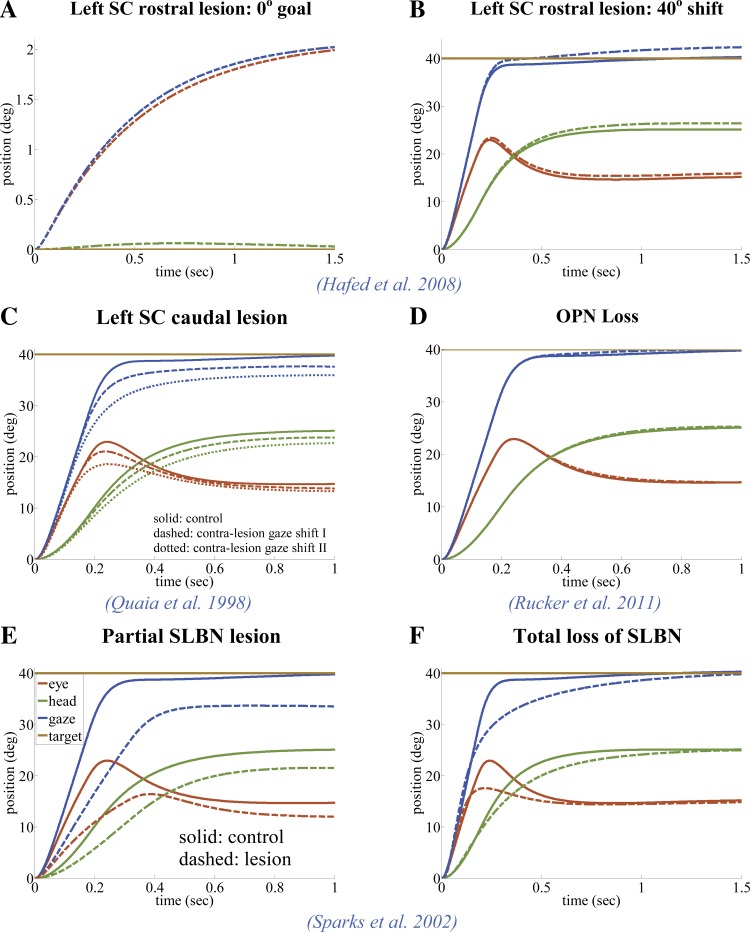
Fig. 10.
Gaze shifts in the SMF model can stop despite focal lesions in key sites (default parameter set, except as specified). Solid lines are control, and dashed or dotted lines are lesion cases; experimental sources are cited in panels. A and B: the SC gain field tv is shifted laterally by ∼2° toward the healthy side: this results in a shift of the null position (target at 0; A) and slightly hypermetric contralateral saccades with the same bias (B). C: unilateral focal lesions in the caudal SC reduce the SC gain field in that region compared with the control case, as well as shifting the tv curve null position, due to their impact on the location of minimal projection strength. The result is slower hypometric gaze shifts in the contralesional direction (consistent with Quaia et al. 1998). Two exemplary lesion cases are illustrated in this figure: case II (dotted lines) represents a more severe caudal lesion compared with case I (dashed lines). The distinction between A and C is that in A the lesion is inside the rostral zone, which corresponds to the retinal areas represented strongly by both colliculi, whereas in C the lesioned area corresponds to a visual field represented by one colliculus (see Fig. 5 for the changes in bilateral projection fields from SC). D: loss of OPNs in the circuit forces the response to remain in saccadic mode; hence, the response is the same as the control during the saccade interval, with a small increase in drift speed during the fixation interval due to the unmasked faster dynamics. E: ipsilateral SLBN gain (B) is reduced by 50% throughout, causing slower and hypometric gaze shifts to the ipsilateral side, replicating lidocaine injections in the PPRF. F: total loss of SLBN (acute) results in very slow, but accurate, gaze shifts with the dynamics of the slow mode.

