
Figure 5.
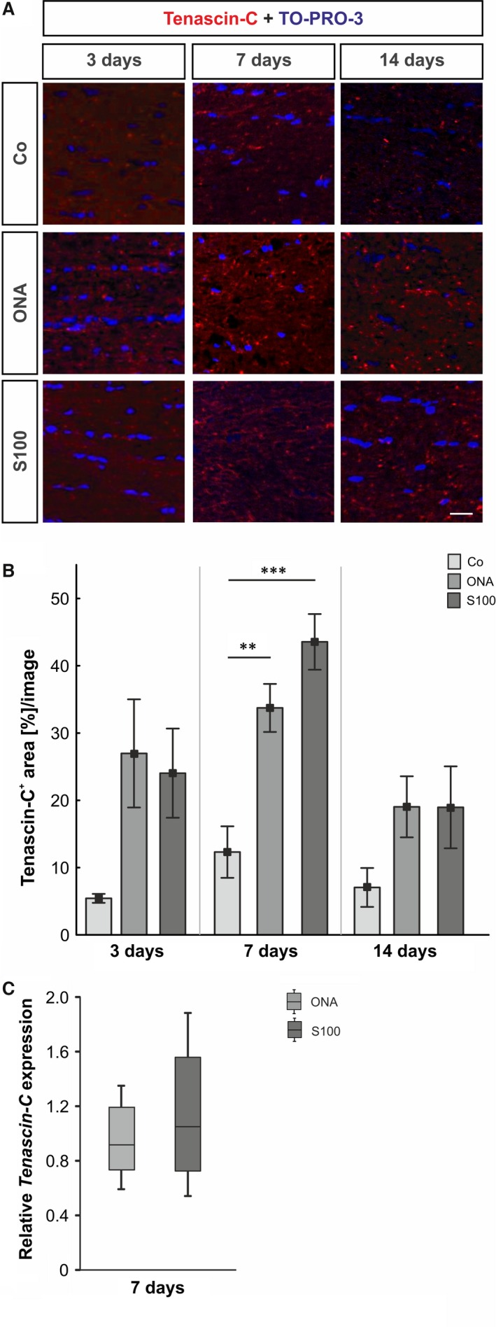
(A) Optic nerves were stained with an anti‐tenascin‐C antibody (red) at 3, 7 and 14 days. Cell nuclei were visualized with TO‐PRO‐3 (blue). (B) The immunoreactivity of tenascin‐C did not change in both groups after 3 days (P > 0.05). At 7 days, an up‐regulation of tenascin‐C was noted in the ONA group (P = 0.009) and in the S100 group (P = 0.0009). No alterations could be noted in both groups after 14 days (P > 0.05). (C) The expression of the tenascin‐C mRNA revealed no changes in either group at day 7 (P > 0.05). Values for immunostaining are mean ± S.E.M. and for qRT‐PCR median ± quartile ± maximum/minimum; scale bar: 40 μm; ** P < 0.01, *** P < 0.001.