Figure 1.

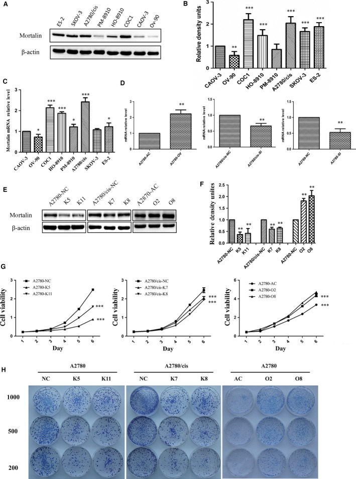
Mortalin is up‐regulated in ovarian cancer cell lines and promotes ovarian cancer cell growth. (A) Western blot analysis using anti‐mortalin antibody to evaluate mortalin (75 kD) expression in ovarian cancer cell lines. (B) Quantitative results from Western blots. β‐actin was used as internal control. (C) Mortalin mRNA expression in ovarian cancer cells was analysed using Quantitative RT‐PCR. (D) To confirm the transfection efficiency, mortalin mRNA expression in different transfected cells was analysed through Quantitative RT‐PCR analyses. (E) Western blot showed mortalin protein expression in different groups. Reduced mortalin expression in stable knockdown clones generated by shRNA (K5 and K11 of A2780, K7 and K8 of A2780/cis). Scrambled cells (A2780‐NC and A2780/cis‐NC) were the non‐specific shRNA control groups. Stable mortalin‐expressing clones (O2 and O8) were established in A2780 cells, Control cells (A2780‐AC) were transfected by empty vectors. (F) Quantitative results from Western blots. β‐actin was used as a loading control. (G) CCK‐8 cell proliferation assay demonstrated that mortalin overexpression in A2780 cells induced significantly higher growth rates compared with their vector controls. In contrast, mortalin depletion in A2780 and A2780/cis cells remarkably decreased cell proliferation compared with their scrambled controls. (H) Colony formation assay showed that mortalin‐overexpressing cells formed much larger clones. *P < 0.05, **P < 0.01, ***P < 0.001.
