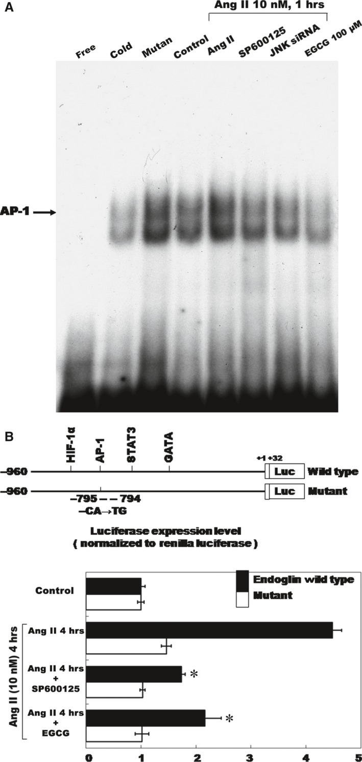
Figure 4.

Angiotensin II‐endoglin expression increased the AP‐1‐binding activity and EGCG inhibits promoter activity. (A) Representative electrophoretic mobility shift assay showing protein binding to the AP‐1 oligonucleotide in the nuclear extracts of CFs following Ang II induction both in the presence and in the absence of either EGCG (100 μM) or JNK1 inhibitor (SP600125). Similar results were observed in two other independent experiments. A significant supershifted complex (super) following the incubation with AP‐1 antibody was observed. Cold oligo means unlabelled AP‐1 oligonucleotides. (B) Constructs of AP‐1 promoter gene. Positive +1 demonstrates the initiation site for the endoglin transcription. Mutant endoglin promoter indicates mutation of AP‐1‐binding sites in the endoglin promoter as indicated. Quantitative analysis of the resistin promoter activity. Cultured CFs were transiently transfected with the pAP‐1‐Luc by gene gun. The luciferase activity in cell lysates was measured and normalized with the renilla activity (n = 3 per group). *P < 0.05 versus control. CFs, cardiac fibroblasts; EGCG, epigallocatechin‐3‐O‐gallate.
