Figure 2.

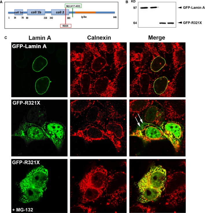
Expression and localization of GFP‐R321X in HEK293 cells. (A) Localization of the nonsense mutation (R321X) upstream of the nuclear localization sequence (NLS) on the Lamin A/C protein. (B) Lysates from HEK 293 cells expressing either GFP‐Lamin A or GFP‐R321X were subjected to Western blot analysis using a monoclonal antibody raised against GFP. (C) HEK293 cells were transfected with either GFP‐Lamin A (upper panel) or GFP‐ R321X (middle and lower panel) and analysed after 24 hr by confocal laser‐scanning microscopy using a polyclonal anti‐calnexin antibody (red) as ER marker. Colocalization is shown in yellow in the merge panels. Note the marked differences in Lamin A distribution in cells expressing R321X, which accumulates within the ER and the nucleoplasm compared with the normal nuclear staining in cells expressing GFP‐Lamin A. White arrows in the Merge panel indicate sites of R321X accumulation outside the ER that disappeared after pre‐treatment with the proteasome inhibitor MG‐132 (lower panel).
