
Figure 1.
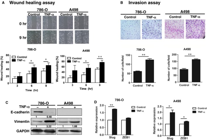
TNF‐α enhanced migration, invasion and EMT of RCC cells. (A) 780‐O and A498 cells were scratched and treated with or without TNF‐α (50 ng/ml) for the indicated times. Cell migration was quantified. (B) 786‐O and A498 cells were treated with or without TNF‐α (50 ng/ml) for 3 days, and the invasion ability was examined. (C, D), 786‐O and A498 cells were treated with or without TNF‐α (50 ng/ml), and the EMT markers were examined by Western blot (C) and RT‐qPCR (D). The results are representative of three independent experiments. *P < 0.05; **P < 0.01. EMT, epithelial‐mesenchymal transition; TNF‐α, tumour necrosis factor‐alpha.