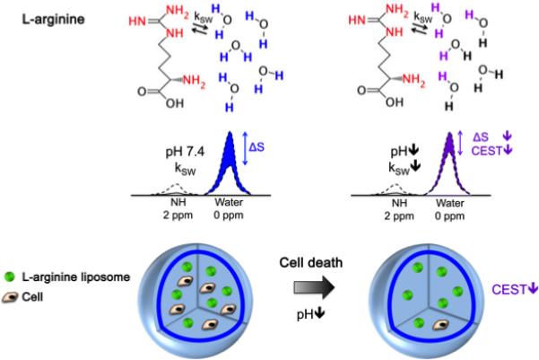
Figure 6. Schematic showing the principles of in vivo detection of cell viability using LipoCEST microcapsules as pH nanosensors.

The CEST contrast is measured by the drop in the signal intensity (ΔS) of water after selective saturation (i.e. removal of capability to generate signal) of the NH protons in L-arginine at 2 ppm. The L-arginine protons (red) inside the LipoCEST capsules exchange (kSW) with the surrounding water protons. The kSW is reduced at lower pH causing a significant drop in CEST contrast. Reproduced from (9) with permission.
