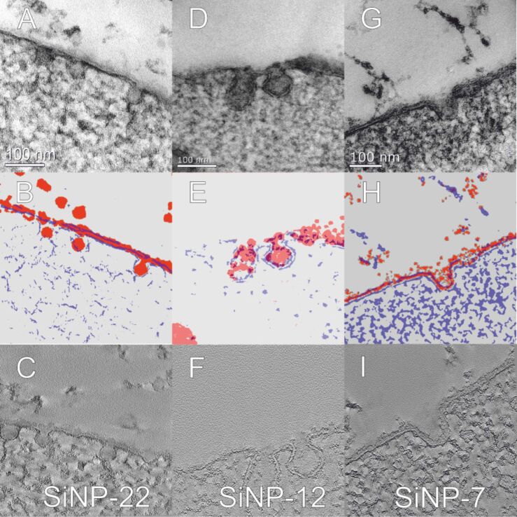
Figure 11.
TEM brightfield micrographs of high-pressure frozen hMSC cells exposed to SiNPs at a concentration of c = 75 µg·mL−1 for 10 min, stained with OsO4 and uranyl acetate (micrographs A, D and G show SiNP-22, SiNP-12 and SiNP-7, respectively). Prior and during the incubation, the hMSCs were kept at 4 °C. For clarification purposes the illustrations below (B, E and H) represent the membrane (blue) and the SiNPs (red) as extracted from energy filtered TEM measurements of the above micrographs. Finally, the micrographs in the bottom row show an optical slice from the respective tomography reconstruction (C, F and I).

