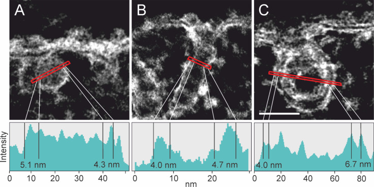
Figure 5.
STEM micrographs showing the particle surrounding membrane formed upon uptake of SiNP-22 (A), SiNP-12 (B) and SiNP-7 (C), respectively. The thickness of the membrane is measured from the intensity profile (below). Membrane thickness varies little and is found to be between 4 and 7 nm for all three different particle sizes. For SiNP-22 and SiNP-12 the typical contrast characteristic for a double layer membrane is unincisive as can be seen in the respective intensity profiles. Experimental conditions same as in Figure 4. Scale bar = 50 nm.

