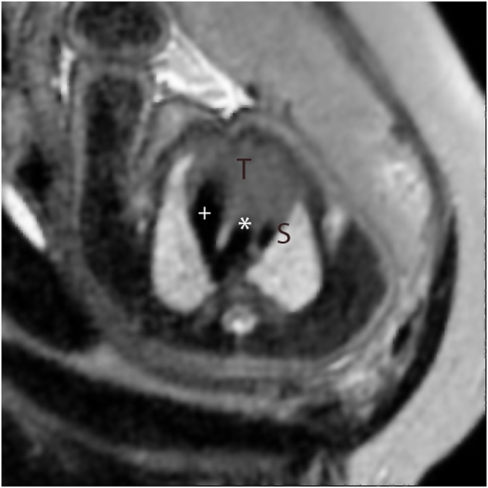
Figure 2.

Single‐shot fast spin‐echo (SSFSE) black‐blood image in a high transverse orientation in a normal fetus. This corresponds to the standard ‘three‐vessel view’ in fetal echocardiography showing the V‐shaped connection between arterial duct (+) and the aorta (*) adjacent to the superior caval vein (S). The thymus gland is seen anteriorly (T)
