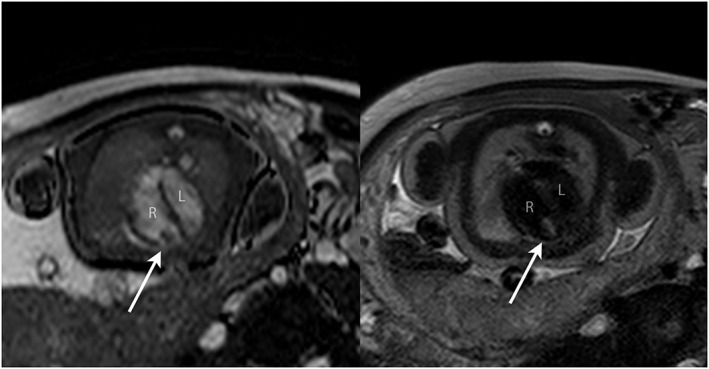
Figure 8.

Balanced SSFP bright blood image (A) and single‐shot fast spin‐echo (SSFSE) black‐blood image (B) in a 38‐week fetus with an RV mass (arrowed). Based on the size, position and tissue signal on multiple sequences, a solitary ventricular rhabdomyoma was suspected; no other masses were seen including on concomitant fetal brain MRI. All findings were confirmed postnatally. R, right ventricle; L, left ventricle
