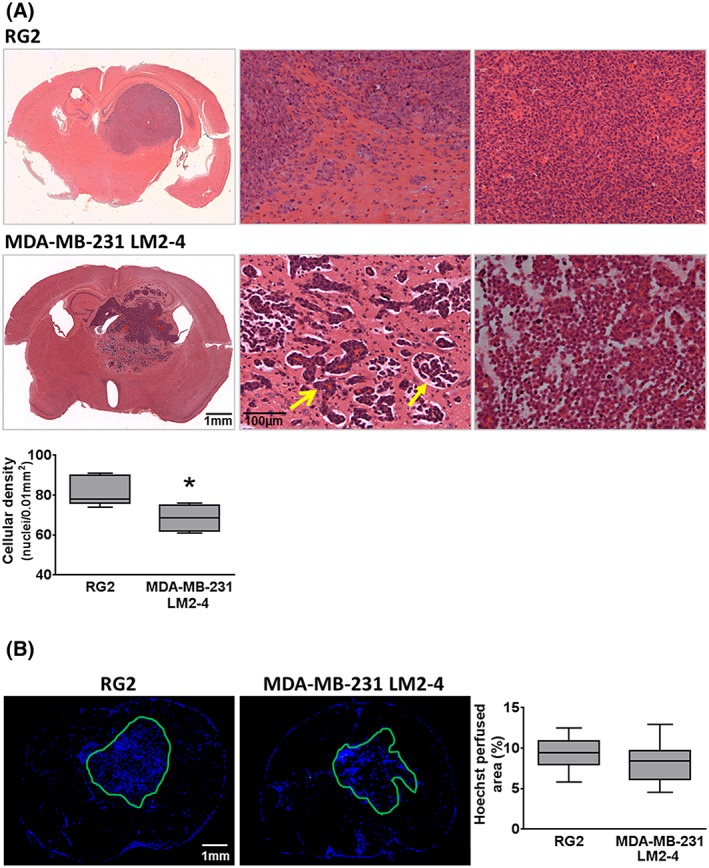
Figure 2.

A, H&E staining of RG2 and MDA‐MB‐231 LM2–4; whole brain composite images and 100× images of the tumour periphery (middle panel) showed RG2 tumours as relatively well circumscribed masses with some local invasion at the periphery, and MDA‐MB‐231 LM2–4 tumours as substantially locally invasive (open head arrow shows tumour cells surrounding a blood vessel) with associated oedema (closed head arrow). The right‐hand panel shows cell density at the centre of the tumours. Mean cellular density was assessed in both tumour types; MDA‐MB‐231 LM2–4 tumours were significantly less dense. B, Fluorescence microscopy of Hoechst 33342 uptake in representative RG2 and MDA‐MB‐231 LM2–4 tumour bearing brains revealed no significant difference between the perfused areas in the two tumour types. *p < 0.05, unpaired Student's t‐test
