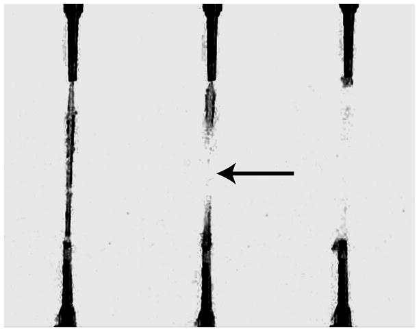
Figure 3. Visual Confirmation of a Tensile Break.

Three frames (captured at 30 frames per second) from a video of uniaxial tensile test are shown. The left frame shows the gel just before its tensile failure. The middle frame confirms a tensile break in the center of the fibrin gel (arrow) with the two halves of the fibrin gel recoiling back to their respective VAA grips (right frame).
