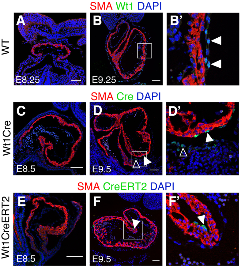
Figure 6.
Cardiomyocyte recombination does not correlate with Cre or CreERT detection in cardiomyocytes (A,B) show immunofluorescence with anti-Wt1 and anti-SMA antibodies on sections of the developing cardiac tube of wild type embryos. (B’) shows a high magnification from the boxed area in (B). Arrowheads indicate pericardial Wt1+ cells. (C,D) show immunofluorescence with anti-Cre and anti-SMA antibodies on sections of the developing heart tube of Wt1Cre embryos. (D’) shows specific areas magnified from (D). Solid arrowheads indicate Cre-positive epicardial cells; empty arrowheads indicate Cre-positive pro-epicardial cells. (E,F) show immunofluorescence with anti-ER and anti-SMA antibodies on sections of the developing heart tube of Wt1CreERT2 embryos. (F’) shows specific areas magnified from F. Solid arrowheads indicate CreERT2-positive epicardial cells. Bar is 50 μm in (A,B) and 100 μm in (C–F).

