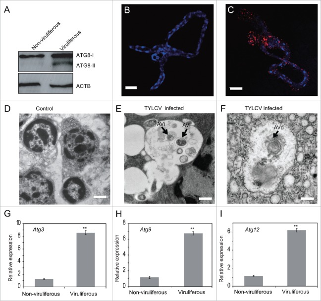
Figure 1.
TYLCV infection induces autophagy in whiteflies. (A) Immunoblot analysis of whiteflies infected with TYLCV for 24 h and transferred to cotton for 120 h. ATG8-I (16 kDa) is observed in both viruliferous and nonviruliferous whiteflies, and ATG8-II (14 kDa) is induced only in the viruliferous whiteflies. Midguts of nonviruliferous (B) and viruliferous (C) whiteflies were fixed and immunofluorescence labeled with anti-ATG8 antibody and secondary antibody conjugated to Dylight 549 (red). Blue indicates DAPI staining of the nuclei. Twenty midguts of viruliferous whiteflies were measured and 95% of them were positive. A representative image is shown, Bar: 50 μm. TYLCV infection induces autophagosome formation as measured by electron microscopy (D to F). Representative images are shown for nonviruliferous (D, Bar: 1 μm) and viruliferous whiteflies (E and F, Bar: 0.5 μm). The initial autophagic vacuole (AVi)/autophagosome can be identified by its rough endoplasmic reticulum, and a double membrane (E). The multimembrane structure in the degradative autophagic vacuole (AVd)/autolysosome can be observed as well (F). Relative expression level of Atg3 (G), Atg9 (H) and Atg12 (I) were tested by qRT-PCR and ACTB was used as the internal control (*, P < 0.05; **, P < 0.01).

