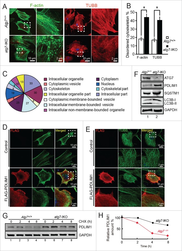
Figure 4.
PDLIM1 is degraded via an ATG7-mediated autophagy-lysosome pathway to maintain the proper organization of the cytoskeleton. (A) IF of F-actin and TUBB in Atg7+/+ (upper panel) and atg7-IKO (lower panel) MEFs. The enlarged images originated from the dotted squares. (B) The percentage of disordered F-actin and TUBB cytoskeleton in (A). (C) Cluster analysis of upregulated proteins in atg7-IKO MEFs. (D, E) Co-IF of F-actin (D) and TUBB (E) with overexpressed FLAG-PDLIM1 in MEF cells, Framed area was magnified. Note that the overexpression of PDLIM1 impaired the disorganization of cytoskeleton. (F) Immunoblotting analysis of ATG7, PDLIM1, SQSTM1 and LC3B in both Atg7+/+ and atg7-IKO MEFs. (G) Cycloheximide (CHX) chase assay of PDLIM1 in Atg7+/+ and atg7-IKO MEFs. Samples were taken at 0, 2, 4, 8 h after the addition of CHX and PDLIM1 level was tested by immunoblotting. (H) Quantification of PDLIM1 level in the CHX assay using the ratio of PDLIM1:GAPDH.

