
Figure 5.
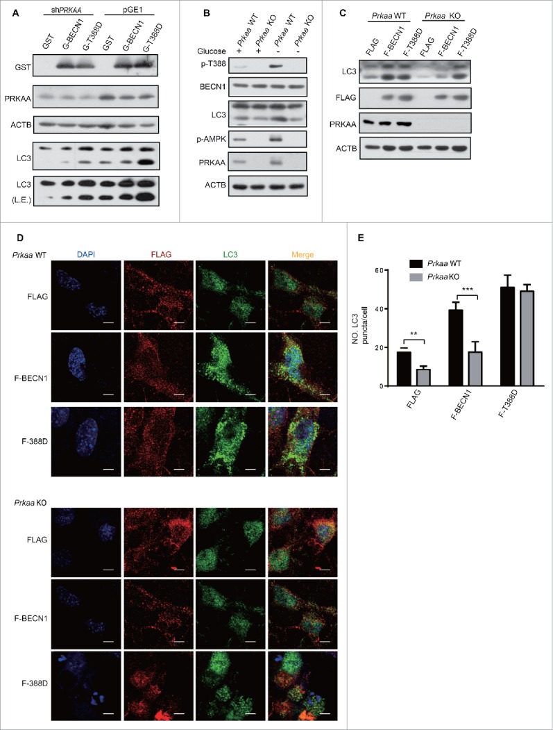
AMPK is necessary for BECN1 Thr388 phosphorylation-induced autophagy. (A) PRKAA knockdown decreased autophagy in cells transfected with GST and GST (G)-BECN1. After coexpression of pGE1 or pGE1-shPRKAA and GST, GST-BECN1WT or GST-BECN1T388D in HEK293T cells for 48 h, cell lysates were probed with the indicated antibodies. L.E., long exposure. (B) Decreased BECN1 T388 and attenuated autophagy were found in Prkaa KO MEF cells. Prkaa WT and KO MEF cells were subjected to glucose starvation for 3 h. p-BECN1 and LC3-II were detected. (C) Autophagy enhanced by phosphorylation of BECN1 is specifically detected in Prkaa WT MEF cells. MEFs were infected with lentivirus and lysed after 48 h. F, Flag. (D) Immunofluorescence was performed after MEFs were infected with the indicated virus. Representative images of endogenous LC3 puncta are shown as indicated. Scale bars: 10 μm. (E) Quantification of GFP-LC3 puncta. Bars are mean ± SEM of triplicate samples (≥ 10 cells analyzed per sample).