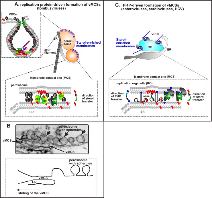
Fig 1. VMCSs promote the morphogenesis and functioning of viral replication organelles.
(A) The formation of invagination-type replication organelles is driven by vMCSs through direct protein–protein interactions of a viral replication protein (e.g., TBSV p33) with cellular ORPs and the ER residential VAPs. The vMCS, which is likely stabilized by co-opted actin filaments, facilitates the enrichment of sterols in the peroxisomal membrane. (B) An electron microscopic (EM) image of a vMCS and multiple vesicle-like structures (spherules), each of which harbors a viral replicase complex (VRC), which replicates the viral genome, is shown from a TBSV-infected plant cell [13]. We propose that the vMCS forms transiently between two subdomains of apposing membranes, locally enriching sterols, then sliding onto new neighboring subdomains of the same organelles, thus acting as a molecular assembly line. (C) vMCSs between protrusion-type replication organelles and the ER depend on the recruitment of PI4Ks by VPs. At the replication organelle membranes, PI4Ks produce PI4P lipids to recruit OSBP. Then, OSBP mediates the accumulation of cholesterol in replication organelle membranes in a counter-exchange with PI4P, which is hydrolyzed after delivery at the ER by Sac1 phosphatase [10]. OSBP is naturally anchored to the ER by VAPs, which for some viruses are known to interact with VPs (see text).

