
Figure 3. Dissociated hDRG neurons over time in vitro.
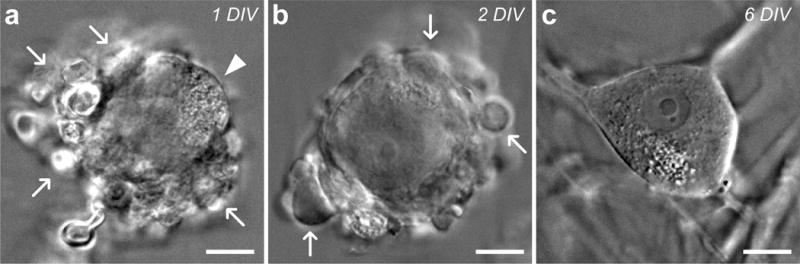
Infrared differential interference contrast microscopy (IR-DIC) images of cultured human sensory neurons. (a) Initially, most dissociated neurons are encased in glial cells (white arrows) after 1 day in vitro (DIV), but can be identified by partially visible plasma membrane (white arrowhead). (b) As time in culture progresses, glia (white arrows) continue to peel off and adhere to the coverslip, exposing more of the plasma membrane. (c) After 6 DIV, the plasma membrane of most neurons is completely exposed, leaving them amenable to patch-clamp recordings and calcium imaging studies. Scale bars represent 20 μm.