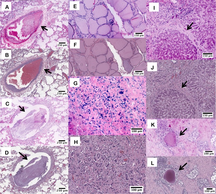
Fig 2. D&E and H&E images from FFPE tissue sections.
D&E (A) and H&E (B) of lung parenchyma and small pulmonary artery branch with blood clot (arrow). D&E (C) and H&E (D) of bronchus with mucus plug (arrow). D&E (E) and H&E (F) of thyroid follicles. D&E (G) and H&E (H) of cirrhotic liver showing ductular reaction. D&E (I) and H&E (J) of liver with a cirrhotic nodule (arrow) and surrounding ductular reaction. D&E (K) and H&E (L) of prostate glands with corpora amylacea (arrow).

