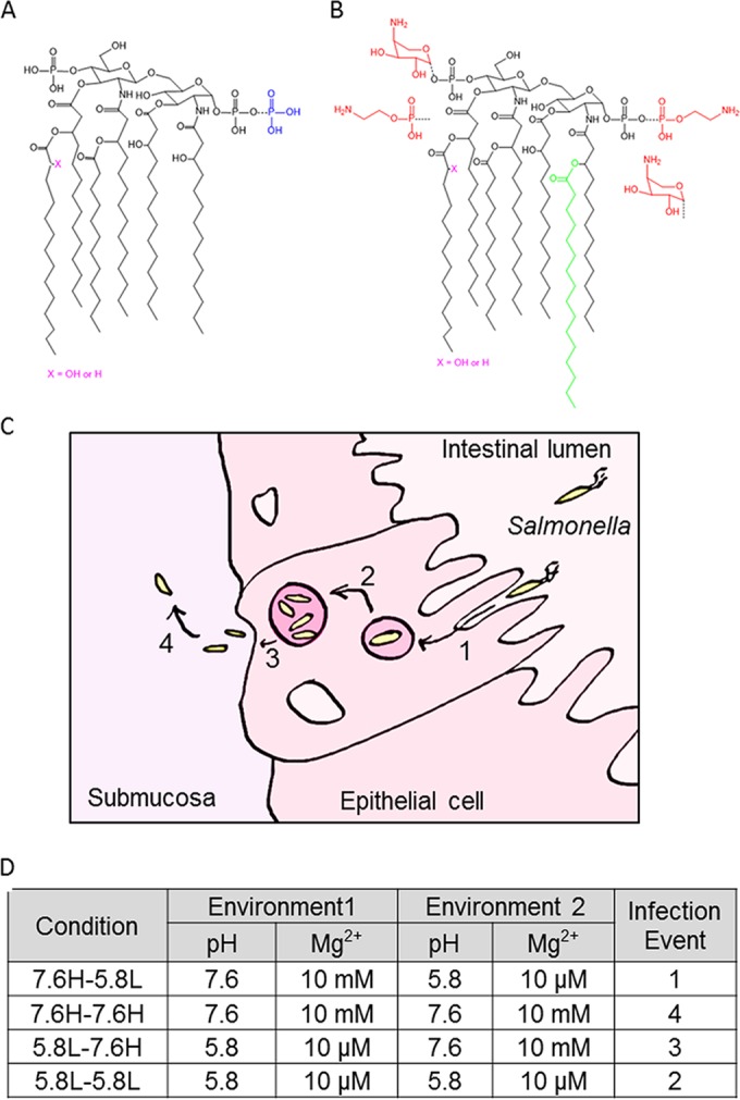
FIG 1 .

Salmonella lipid A structures present under different environmental conditions. The colored additions (blue, additional phosphate; red, pEtN and l-Ara4N; green, palmitoyl chain) to the base structure are representative of variations present at pH 7.6 10 mM Mg2+ (A) and pH 5.8 10 µM Mg2+ (B). (C) Diagram indicating the infection events mimicked by the environmental shift conditions. Salmonellae invade epithelial cells and become contained within an acidifying vacuole in step 1. Step 2 is Salmonella replication within this acidic SCV. The third step is Salmonella release into the submucosa, while the fourth step is survival and dissemination in this location. (D) The various environmental shift protocols are listed, with the high Mg2+ concentration corresponding to 10 mM MgSO4 and the low Mg2+ concentration corresponding to 10 µM MgSO4.
