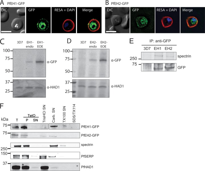
FIG 2 .
PfEH1 and PfEH2 interact with the erythrocyte cytoskeleton. IFA of paraformaldehyde/glutaraldehyde-fixed, trophozoite/early schizont-stage parasites expressing PfEH1-GFP (A) or PfEH2-GFP (B) under control of the respective endogenous promoter. The GFP-expressing parasites were costained with anti-RESA (red). The nuclei were stained with DAPI (blue). Bars = 5 µm. DIC, differential interference contrast. (C and D) Western blots of PfEH expression in trophozoite-stage parasites. PfEH proteins are not detected in 3D7 parent parasites, barely detected when GFP tagged at the endogenous locus (endo), and highly expressed in the episomal overexpression (EOE) lines. P. falciparum haloacid dehalogenase 1 (PfHAD1) is used as a loading control. α-GFP, anti-GFP antibody. (E) Immunoprecipitation (IP) of RBCs infected with the 3D7 parent line, PfEH1-GFP or PfEH2-GFP (EOE lines). Complexes were isolated with anti-GFP antibodies, and the final eluate was probed with anti-GFP antibody to confirm pulldown of the PfEH proteins and with human antispectrin antibody. The positions of molecular mass markers (in kilodaltons) are shown to the left of the blots in panels C to E. (F) Sequential fractionation of the 3D7 parent line, PfEH1-GFP, or PfEH2-GFP (EOE lines). The RBC plasma membrane was permeabilized with tetanolysin O (TetO). The total (T) fraction, pellet (P) fraction, and supernatant (SN) fractions are shown in the three leftmost portion of the blots. The pellet fraction was then subjected, sequentially, to hypotonic lysis (Tris HCl) to release soluble parasitophorous vacuole (PV)/parasite/Maurer’s cleft contents, carbonate (Carb.) to release membrane fraction-associated contents, Triton X-100 (TX100) to release membrane contents, and the final pellet was solubilized in Triton X-114 (TX114)/SDS. P. falciparum serine-rich protein (PfSERP) is a soluble parasitophorous vacuole integrity marker, and PfHAD1 is a soluble parasite integrity marker.

