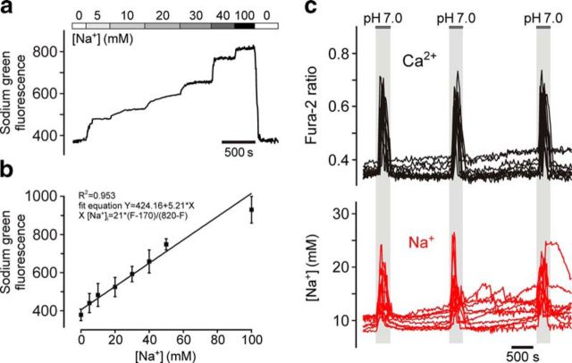
Figure 3.
Estimation of absolute resting level and peak acidification-induced increases in [Na+]i in chemosensitive brainstem astrocytes. a, Calibration of Na+-sensitive fluorescence signal in astrocytes of organotypic brainstem slices recorded in the presence of gramicidin D, monensin, and ouabaine (to equilibrate Na+ across the cell membrane) at increasing extracellular [Na+]. Averaged trace of Sodium Green fluorescence changes in 152 astrocytes recorded in three organotypic brainstem slices is shown. b, Sodium Green fluorescence plotted as a function of [Na+]. c, Representative example illustrating the resting level and peak acidification-induced increases in [Na+]i in chemosensitive brainstem astrocytes. Traces depict responses of 10 individual cells recorded in an organotypic brainstem slice preparation.

