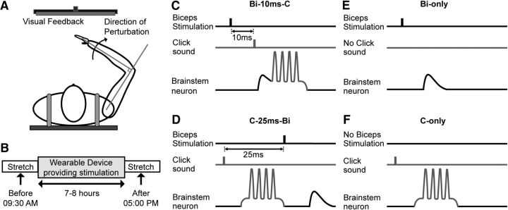
Figure 1.
Schematic diagram showing the experimental setup and wearable device stimulus conditions. A, Subjects were strapped into a chair, with their right arm attached to a robotic device capable of delivering extension perturbations at the elbow joint. A computer screen provided visual feedback of the elbow angle. B, The general experiment protocol. C–F, The four different stimulus conditions implemented by the wearable device, and their hypothesized effects on a reticulospinal neuron. C, Biceps stimulation 10 ms before the click (Bi-10ms-C); the EPSP elicited by the afferent input arrives just before the click-induced discharge, which should potentiate synapses conveying the EPSP. D, Click 25 ms before biceps stimulation (C-25ms-Bi); the afferent EPSP arrives after the click-induced discharge, which should lead to depression of the EPSP. E, Biceps stimulation alone (Bi-only). F, Click stimulation alone (C-only). With no stimulus pairing, we expect no change in EPSP amplitude.

